Laser removal is considered when a stone is unlikely to pass, keeps causing pain or blockage, threatens kidney function, or sits behind an infection that must first be controlled. You will learn how stone size, location, composition, test results, and treatment risks determine whether observation, medication, shock-wave treatment, ureteroscopy, or PCNL is the safer choice.
Key takeaways
- Choose active removal for obstruction, kidney damage, uncontrolled pain, or infection risk.
- Stone size, location, density, and composition guide treatment selection.
- CT imaging and kidney function tests help confirm whether laser ureteroscopy fits.
- Ask how laser ureteroscopy compares with shock waves and PCNL for your stone.
When waiting for a kidney stone is no longer the safer plan
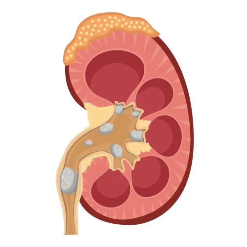
Persistent kidney stone pain, recurrent renal colic, or pain that medication cannot control makes active removal preferable to continued observation. The same applies when imaging shows persistent urinary obstruction, worsening hydronephrosis, reduced kidney function, bleeding, stone growth, or a stone too large to pass naturally.
| Choice | When waiting is reasonable | When active removal is preferable |
|---|---|---|
| Observation | Small, asymptomatic, nonobstructing kidney stone; reliable imaging follow-up | Growth, new symptoms, obstruction, or uncertain follow-up |
| Medication-assisted passage | Selected small ureteral stone, especially near the bladder, with no infection, kidney-function decline, or uncontrolled pain | Failure to pass, repeated emergency visits, persistent pain, or ongoing blockage |
| Ureteroscopic laser treatment | Not usually needed for a stone likely to pass naturally | Unfavourable size or location, persistent obstruction, or failed observation or medication |
| PCNL | Not used for routine small stones | Stone burden larger than about 2 cm, where multiple ureteroscopic procedures may be needed |
An obstructed kidney with fever, chills, infection symptoms, or sepsis requires urgent drainage with a ureteral stent or nephrostomy and antibiotics. Do not treat the stone definitively with laser while infection remains uncontrolled; fragmentation follows after the infection resolves.
A small, nonobstructing renal stone can be monitored because it may cause no immediate harm. That plan becomes unsafe when follow-up imaging shows obstruction or growth, urine tests suggest infection, blood tests show declining kidney function, or symptoms continue despite appropriate medication.
How size, location, and composition determine the treatment choice
A 4-mm stone near the bladder has a reasonable chance of passing, while a 15-mm stone in the kidney usually needs active removal. There is no universal kidney stone size threshold: the decision also depends on ureter stone location, obstruction, symptoms, anatomy, and the likelihood that fragments will clear.
| Option | What the measurement or characteristic suggests | Typical role |
|---|---|---|
| Observation | Small, uncomplicated ureter stone, especially under 5 mm | Wait for passage with imaging follow-up when pain, infection, obstruction, and kidney function remain controlled |
| Medical expulsive therapy | Selected distal ureter stones, often 5–10 mm | An alpha-blocker may assist passage; it is not a substitute for treatment when obstruction or infection persists |
| Shock-wave lithotripsy | Smaller kidney or ureter stones with favorable location and lower density | Least invasive stone-breaking option, but fragments may require repeat sessions |
| Laser ureteroscopy | Larger, hard, obstructing, or poorly positioned stones | Direct fragmentation and removal; often favored for hard stones or when one procedure is important |
| PCNL | Large renal burden, commonly over 20 mm, or staghorn stones | Percutaneous removal of substantial stone volume, with greater bleeding and recovery considerations |
A lower-pole kidney stone over 1 cm is harder to clear after shock waves because fragments settle below the kidney’s drainage angle; flexible ureteroscopy with laser or PCNL is commonly considered instead. Stone density and composition also matter: high-density calcium oxalate monohydrate or cystine stones resist shock waves, making laser fragmentation more attractive.
What tests confirm whether laser treatment is appropriate
A non-contrast CT for kidney stones shows the stone’s exact size, location, density, and effect on urine drainage. It can reveal high-grade obstruction that makes waiting unsafe. An ultrasound kidney stone evaluation avoids radiation and can show hydronephrosis, but it can miss small ureteral stones; use it first during pregnancy.
Urine testing determines whether laser treatment is safe today. A urinalysis can show blood, white cells, or nitrites, while a urine culture identifies bacteria and guides antibiotic selection. Fever, chills, fast heart rate, low blood pressure, or pus in the urine raise concern for infection behind a blockage.
Blood testing adds information that imaging cannot provide:
- A complete blood count can show an elevated white-cell count associated with infection.
- A creatinine kidney function test shows whether obstruction has impaired filtration.
- Electrolyte testing detects chemical disturbances that affect anaesthesia and urgent care.
An infected, obstructed urinary system is a urologic emergency. Do not fragment the stone immediately: urgent drainage with a ureteral stent or nephrostomy, plus appropriate antibiotics, comes first. Laser removal usually waits until the infection has resolved.
Pregnancy changes both the imaging plan and the treatment threshold. Use ultrasound initially; avoid shock-wave lithotripsy. Ureteroscopy with laser can be considered for obstruction, infection, or unmanageable symptoms after coordination with obstetric and anaesthesia teams, while nonurgent treatment is deferred when possible.
What happens during laser ureteroscopy, and will one procedure be enough
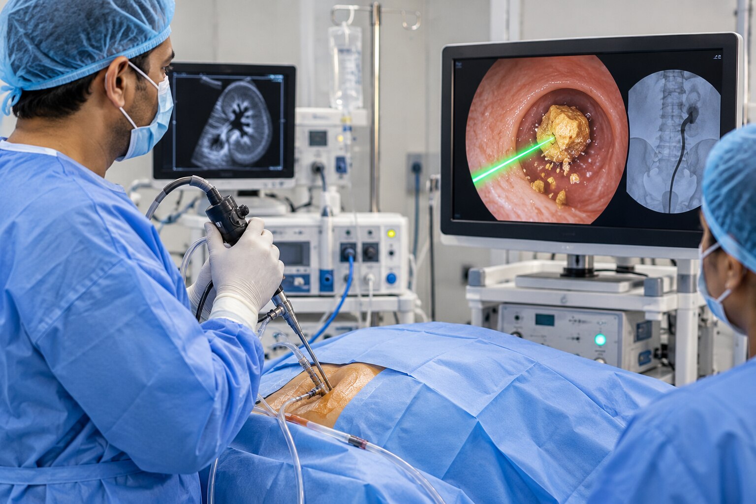
The surgeon guides a thin camera through the urethra and bladder into the ureter or kidney, then uses laser energy to break the stone during a laser lithotripsy procedure. Flexible ureteroscopy allows access to difficult kidney locations, including the lower pole.
1. The surgeon identifies the stone and directs the laser at it under direct vision.
2. Larger fragments are removed with a small basket when retrieving them is practical. With stone dusting and basket extraction, the surgeon may remove selected pieces while reducing the rest to fine particles that can pass in urine.
3. A ureteral stent after ureteroscopy may keep urine draining while swelling settles, especially after a difficult procedure, ureteral injury, infection, or a large stone burden.
A stent is not the treatment itself. Urgency, frequent urination, flank discomfort, and blood in the urine are common while it remains, and you need a documented removal or exchange date; a forgotten stent can cause serious damage.
One procedure is less likely to be enough when the stone is large, hard, numerous, difficult to reach, or when swelling limits access. The surgeon may place a stent first and complete a staged procedure after the ureter relaxes.
Follow-up imaging with ultrasound, an X-ray, or CT checks for residual fragments, obstruction, and silent swelling. Stone analysis and metabolic testing can guide prevention, because “stone-free” does not prevent a new stone from forming.
How laser ureteroscopy compares with shock waves and PCNL
For a ureteral stone, laser ureteroscopy usually offers a higher stone-free rate after one procedure than shock-wave treatment, but it requires anaesthesia and instruments to pass through the urinary tract. The right choice depends on stone burden, location, density, bleeding risk, and your tolerance for repeat treatment.
| Option | Practical advantages | Main trade-offs |
|---|---|---|
| Laser ureteroscopy | Directly fragments and removes stones; effective for hard stones and lower-pole stones; often one procedure | Usually requires general anaesthesia; temporary stent discomfort; risks include infection, bleeding, ureter injury and scarring risks |
| Shock-wave lithotripsy | No urinary-tract instrumentation; lighter anaesthesia and quicker initial recovery | Fragments may remain, especially in the lower pole; hard stones respond poorly; repeat sessions are more likely |
| PCNL | Highest effectiveness for a large kidney-stone burden; percutaneous nephrolithotomy for stones over 2 cm is commonly preferred | General anaesthesia, a small kidney puncture, longer recovery, and greater bleeding-management concerns |
Ureteroscopy is often considered when you cannot stop anticoagulant or antiplatelet medicine, because shock waves and PCNL create more bleeding concerns. A stent can cause urgency, frequent urination, flank discomfort, and blood in the urine; it must be removed or exchanged on schedule.
At Dr. Vivek Jadhav Laser Stone and Urology Clinic, ask which option fits your CT-measured stone size, density, and location, and whether the expected benefit outweighs ureter injury, infection, bleeding, or a second procedure.
Related services
LASER AND COMPLETE ENDOSCOPIC TREATMENT OF URETERIC STONES LASER AND COMPLETE ENDOSCOPIC TREATMENT OF URETERIC STONES Ureteric or Ureteral stones are the kidney stones that get stuck in one or both ureters. View service → |
Laser and endoscopic treatment of kidney stone Laser and endoscopic treatment of kidney stone A complete endoscopic prostate surgery typically refers to a minimally invasive procedure known as transurethral resection of the prostate (TURP). View service → |
Frequently asked questions
When is waiting for a kidney stone no longer the safer plan?
Active removal becomes preferable when pain persists or medication fails, imaging shows obstruction or worsening hydronephrosis, kidney function declines, bleeding occurs, the stone grows, or it is too large to pass naturally.
How do stone size, location, and composition affect laser treatment?
Size and location affect whether a stone can pass or be reached with an instrument. Composition and CT density help predict how easily it will fragment and whether shock waves or another procedure may be more suitable.
What tests confirm whether laser ureteroscopy is appropriate?
Evaluation commonly includes urine testing, blood tests for kidney function, and imaging such as ultrasound or a non-contrast CT scan. The results show obstruction, hydronephrosis, stone size, location, and density.
How does laser ureteroscopy compare with shock waves and PCNL?
Laser ureteroscopy reaches the stone through the urinary passage and suits many ureter and kidney stones. Shock-wave treatment breaks stones from outside the body, while PCNL uses a small kidney access tract for larger or complex stones.