
Waiting is reasonable only when a stone is small, symptoms are controlled, infection is absent, and kidney function is safe. You will be able to distinguish a stone suitable for monitored observation from one that needs urgent drainage or planned removal, compare treatment options, and know what follow-up confirms that the stone is truly gone.
Key takeaways
- Seek same-day care for fever, severe pain, vomiting, one kidney, or inability to urinate.
- A stone’s size, location, obstruction, and movement determine whether observation is reasonable.
- Laser ureteroscopy removes stones through the urinary tract without an external incision.
- Confirm clearance with follow-up imaging and prevent recurrence through stone analysis and urine testing.
When is waiting for a stone to pass no longer safe?
Waiting is no longer safe when a stone threatens your life, kidney function, hydration, or ability to urinate. Seek same-day medical assessment for:
- Fever or chills with a suspected stone, confusion, low blood pressure, or appearing acutely ill
- Inability to urinate, persistent vomiting, dehydration, or pain that prescribed medicine cannot control
- A solitary functioning kidney, known chronic kidney disease, worsening kidney-function blood tests, or ongoing obstruction on imaging
An uncomplicated stone means you are clinically stable, have controllable pain, no infection, and no threatened kidney function. An infected obstructed urinary system is different: bacteria are trapped behind the blockage, making this a urological emergency. Antibiotics alone do not reliably clear trapped urine.
Doctors provide urgent drainage with a ureteric stent or nephrostomy tube drainage, then delay definitive stone removal until infection and sepsis have resolved.
Ask for specialist advice sooner during pregnancy or childhood, with recurrent urinary infections, anticoagulant use, limited access to emergency care, or work where sudden pain could endanger you or others. These factors lower the threshold for assessment or planned treatment.
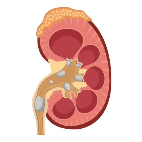
Information about bladder stones does not determine when a kidney or ureteric stone needs removal. Kidney and ureteric stones can obstruct the upper urinary tract and damage kidney function even when bladder symptoms are absent.
How do size, location, and imaging determine whether observation is reasonable?
About 71% of proximal ureter stones under 5 mm pass spontaneously, compared with about 89% of distal ureter stones under 5 mm, according to EAU figures. Rates fall for stones measuring 5–10 mm, and a larger diameter, irregular shape, hard composition, narrow ureter, or unfavourable anatomy further reduces passage.
Stone size and location change the odds:
| Finding | Effect on passage | Practical implication |
|---|---|---|
| Under 5 mm, distal ureter | Highest likelihood | Observation is often reasonable if stable |
| 5–10 mm, especially distal | Lower likelihood | Discuss medical expulsive therapy and follow-up |
| Proximal ureter | Less likely than distal | Allow observation only with closer review |
| Irregular, dense, impacted stone or narrowed anatomy | Reduced likelihood | Planned removal becomes more compelling |
An alpha-blocker such as tamsulosin is most useful for selected distal ureteric stones, particularly those over 5 mm and up to 10 mm. Medical expulsive therapy cannot overcome infection, threatened kidney function, uncontrolled pain, or an impacted stone.
For observation, record the stone’s exact dimensions and location, take prescribed pain relief, and strain urine when advised. Arrange repeat ultrasound or plain radiography when appropriate; use low-dose noncontrast CT when passage or location remains uncertain, or when CT findings show persistent obstruction.
There is no universal waiting period. Stop observing when symptoms recur, obstruction persists, kidney function worsens, or the stone fails to progress. Pain relief does not prove passage: silent obstruction can remain.
What can happen if you keep waiting after the stone stops progressing?
A monitored wait is reasonable only when pain is controlled, infection is absent, kidney function is safe, and follow-up imaging is scheduled. Indefinite postponement is different: a retained or migrating stone can cause recurrent renal colic, hydronephrosis, silent obstruction, repeated emergency visits, infection, work disruption, and repeated analgesic or opioid use.
Persistent obstruction can lead to kidney function loss, emergency admission, or an unexpected urgent ureteric stent or nephrostomy. Pain relief does not prove that the stone has passed.
Active removal becomes more compelling when imaging shows persistent obstruction, the stone grows, symptoms keep returning, the kidney is threatened, or the kidney stone causes infection or significant symptoms.
Stable, asymptomatic renal stones can sometimes be monitored with imaging, but a solitary kidney, chronic kidney disease, recurrent infections, pregnancy, childhood, poor access to emergency care, anticoagulant treatment, or work where sudden pain could endanger people changes the risk calculation.
Ask your clinician:
- What are the stone’s exact dimensions and location?
- Is there hydronephrosis or silent obstruction?
- Are urine culture, urinalysis, creatinine, and other blood tests reassuring?
- How long is observation safe, and what is the consequence of postponing removal?
If symptoms recur or follow-up shows blockage, do not treat “wait and see” as “ignore it.”
Which removal method fits the stone, and what does laser ureteroscopy involve?
The choice depends on stone size, location, hardness, anatomy, kidney function, and your anaesthetic risk—not simply on whether a laser is available.
| Option | What it involves | Main trade-off |
|---|---|---|
| Shock-wave lithotripsy | Shock waves break the stone without entering the urinary tract | Hard stones, larger burdens, deep locations, or unfavourable anatomy can reduce clearance, and repeat sessions may be needed |
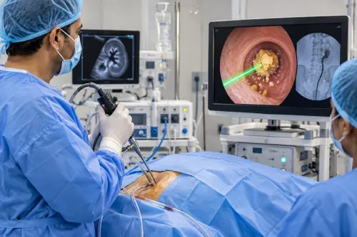
| Ureteroscopy with laser | A thin scope passes through the urethra and bladder into the ureter or kidney; laser fragmentation breaks the stone, followed by basket extraction | Often achieves high clearance in one procedure, but requires anaesthesia and may require a temporary ureteric stent |
| Percutaneous nephrolithotomy | Instruments reach the kidney through a small incision in the back | Generally preferred for renal stone burdens over 20 mm; it is more invasive than the other options |
A stent can cause urinary urgency, frequent urination, flank discomfort, and blood in the urine. Follow-up imaging confirms clearance and checks that drainage has recovered; time away from work depends on the procedure and whether your job involves driving, heavy lifting, or safety-critical tasks.
Laser ureteroscopy is not the first step during sepsis: doctors drain the kidney with a stent or nephrostomy and treat the infection before removing the stone. A large burden may suit percutaneous nephrolithotomy, while unsafe anaesthesia requires stabilisation first.
Dr. Vivek Jadhav Laser Stone and Urology Clinic can help compare these options against your scan, symptoms, and work demands rather than treating one method as suitable for everyone.
Related services
LASER AND COMPLETE ENDOSCOPIC TREATMENT OF URETERIC STONES LASER AND COMPLETE ENDOSCOPIC TREATMENT OF URETERIC STONES Ureteric or Ureteral stones are the kidney stones that get stuck in one or both ureters. View service → |
Bladder Cancer surgery and treatment Bladder Cancer surgery and treatment Bladder cancer surgery typically involves the removal of cancerous tissue from the bladder. View service → |
How do you confirm the stone is gone and reduce the chance of another one?
Reduced pain does not prove passage: a stone can stop hurting while silently obstructing the ureter. Doctors therefore combine symptoms with follow-up imaging, urine tests, and blood tests, including creatinine when kidney function is a concern.
| Test | What it shows | When it fits |
|---|---|---|
| Ultrasound | Remaining stone and whether hydronephrosis has resolved | Follow-up, pregnancy, or radiation avoidance |
| Plain radiography | An X-ray-visible stone and its movement | When the original stone is visible on X-ray |
| Low-dose noncontrast CT | Stone location, size, and obstruction | Uncertain passage or location, balancing radiation exposure |
The choice depends on the stone, pregnancy status, radiation considerations, and original imaging. A temporary ureteric stent also needs a documented removal or exchange date; leaving it untracked creates avoidable complications. After passage or removal, request stone analysis when a fragment is available, a 24-hour urine evaluation when indicated, and blood tests for metabolic causes.
Drink enough to meet your clinician’s advice, then base diet changes on stone type and urine results—not generic restrictions. Kidney stone prevention may target high urinary calcium, excess uric acid, low citrate, infection-related stones, or obstruction causing urinary stasis.
Choose observation only with documented follow-up and emergency instructions. Choose planned removal when passage is unlikely, obstruction persists, symptoms continue, or waiting puts kidney function or patient safety at greater risk.
Frequently asked questions
When is waiting for a kidney stone to pass no longer safe?
Waiting is no longer safe when the stone causes fever or infection, uncontrolled pain, repeated vomiting, dehydration, reduced kidney function, a blocked solitary kidney, or inability to urinate. Seek same-day medical assessment for these warning signs.
How do size, location, and imaging determine whether observation is reasonable?
CT or ultrasound shows the stone’s size, position, degree of obstruction, and effect on the kidney. Small stones low in the ureter are more likely to pass than larger stones lodged higher up, but worsening obstruction or no movement makes observation less suitable.
What can happen if you keep waiting after the stone stops progressing?
A persistent blockage can cause infection behind the obstruction, kidney swelling, declining kidney function, recurrent emergency pain, and difficulty maintaining hydration. Fever with obstruction is an emergency because sepsis can develop.
What does laser ureteroscopy involve?
A urologist passes a small camera through the urinary passage into the ureter, breaks the stone with a laser, and removes or leaves fragments to pass. A temporary ureteral stent can support drainage while swelling settles.
How do you confirm a kidney stone is gone and reduce recurrence risk?
Follow-up imaging confirms whether fragments or obstruction remain. Stone analysis, blood tests, and a 24-hour urine collection identify preventable risks, while adequate fluids, lower salt intake, and targeted dietary changes reduce recurrence.